Redefining Preventive Health with Advanced Imaging
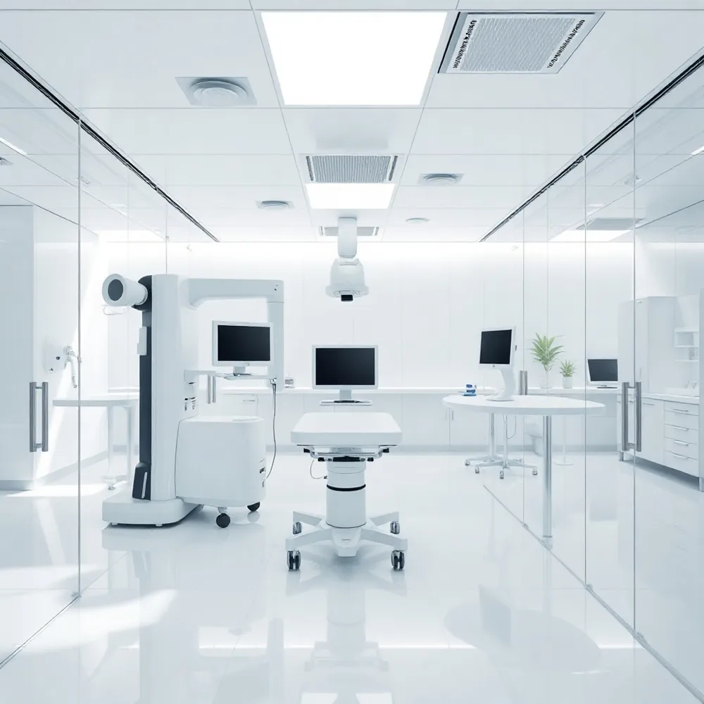
Longevity medicine now treats aging as a modifiable biological process rather than an inevitable decline, shifting the clinical focus from reactive symptom management to proactive intervention. At the Medical Institute of Healthy Aging, we leverage advanced imaging such as Whole-Body MRI to identify preclinical abnormalities before symptoms arise. While some facilities rely on sporadic, symptomatic screenings, our advanced diagnostics provide longitudinal, high-resolution data that tracks systemic age-related health risks with precision. By integrating molecular and structural insights, we translate AI-predicted age differences into personalized longevity pathways.
The Rise of Longevity Clinics: A Proactive Healthcare Model
A longevity clinic is a specialized medical facility that may contribute to extending healthspan by prioritizing preclinical prevention over reactive symptom management. These centers operate as an Analytical Center that functions as the brain of the organization, managing complex data integration from wearables and high-fidelity diagnostic tools. Unlike traditional medicine, which often relies on identifying disease after symptoms emerge, some clinics utilize aging clocks and biomarkers to assess health in individuals who appear practically healthy.
What defines a longevity clinic and its role in personalized medical interventions?
At mdiha.com, patients receive personalized medical interventions rooted in deep phenotyping of the aging process. While some facilities may offer fragmented services, this institution prioritizes a three-level patient routing system that integrates continuous remote monitoring with comprehensive in-clinic diagnostics. These diagnostics often include whole-body MRI and molecular profiling to detect physiological decline years before it manifests clinically.
Data-driven personalization is essential for identifying optimal preventive levers. Multidisciplinary teams at mdiha.com use clustering and time-series analysis to segment patient data into bespoke health plans. By incorporating advanced imaging modalities such as coronary artery calcium scoring and CCTA, these clinicians track health metrics longitudinally. This approach ensures that interventions for metabolic or cardiovascular resilience are continuously refined based on objective, repeatable measures rather than subjective assessments.
Molecular and Physiological Biomarkers of Biological Age
Assessing biological age requires a multifaceted approach that integrates molecular indicators with functional physiological measurements. At the molecular level, epigenetic clocks based on DNA methylation patterns, telomere length assessment, and the analysis of inflammatory cytokine profiles provide critical insights into cellular aging processes. These molecular markers offer a snapshot of systemic inflammaging, allowing clinicians at mdiha.com to track the cumulative impact of cellular senescence over time.
Complementing these molecular data points are functional physiological measurements that evaluate intrinsic capacity. These metrics include cardiorespiratory fitness measured via VO2max, lean muscle mass, grip strength, and gait speed. While some clinics rely solely on blood-based diagnostics, mdiha.com integrates these functional metrics with advanced imaging to capture a more complete picture of systemic resilience. For example, CT body composition analysis can reveal sarcopenia, while brain age estimation via MRI provides a powerful prognostic indicator of future cognitive function.
The synthesis of these diverse data streams is what differentiates a proactive longevity framework from traditional reactive medicine. By combining bone density measurements with real-time molecular markers, clinicians can better quantify an individual's biological departure from their chronological age. This integrated diagnostic framework enables a comprehensive assessment of an individual’s healthspan and supports the implementation of personalized interventions that address the root causes of age-related decline.
Detecting Dementia Early: Neuroimaging and Blood Biomarkers
 Advanced neuroimaging plays a critical role in the early diagnosis and prognosis of dementia by visualizing pathological biomarkers long before clinical symptoms manifest. At mdiha.com, structural MRI is utilized to quantify hippocampal atrophy and cortical thinning, which serve as essential markers for neurodegeneration. Molecular imaging, specifically PET scans using specialized radiopharmaceutical tracers, enables the direct visualization and quantification of amyloid-beta plaques and tau protein tangles in the brain. Furthermore, functional imaging techniques like FDG-PET allow clinicians to assess regional glucose metabolism, identifying patterns of synaptic dysfunction characteristic of various dementia etiologies. When integrated with multimodal data analysis, these imaging modalities are studied for their potential to support precise diagnostics and monitoring of disease progression.
Early detection of Alzheimer's disease now relies on a sophisticated integration of blood-based biomarkers, neuroimaging, and clinical cognitive assessments. Key plasma biomarkers include the Aβ42/Aβ40 ratio, various phosphorylated tau species (specifically p-tau181 and p-tau217), neurofilament light chain (NfL), and glial fibrillary acidic protein (GFAP). A 2024 Nature study highlights how the integration of multimodal imaging with these biological markers improves clinical precision. While elevated levels of these proteins are associated with a significantly increased risk, current research indicates that combining markers may offer improved predictive performance in research settings. Beyond these, a deep learning algorithm applied to brain MRI scans can estimate brain age and identify a Predicted Age Difference (PAD), where a higher score correlates to increased cognitive impairment.
| Diagnostic Tool | Clinical Target | Clinical Value |
|---|---|---|
| Structural MRI | Hippocampal atrophy | Quantifies neurodegeneration |
| Amyloid/Tau PET | Protein pathology | Confirms AD etiology |
| Plasma Biomarkers | p-tau217/GFAP | High-throughput screening |
| AI-driven PAD | Brain age | Predicts decline rate |
Unlocking Longevity Insights with Radiomics and AI
 Radiomics offers a high-dimensional approach to data extraction from standard medical images, such as CT and MRI scans. By using high-throughput computational techniques, radiomics analyzes complex patterns that are otherwise invisible to the human eye, effectively performing a top-down phenotyping of a patient. This method captures the cumulative impact of both genetic predispositions and environmental exposures, offering a more comprehensive assessment than isolated clinical tests. At mdiha.com, we integrate these advanced metrics into our Whole-Body Imaging & Early Detection services to identify latent pathology years before clinical manifestations occur.
Machine learning has fundamentally changed mortality prediction by allowing algorithms to derive insights from routine imaging. Research indicates that deep learning models applied to chest CT scans can achieve a c-index of 0.68 for mortality prediction, a metric comparable to established tools like the Framingham risk model. Unlike traditional diagnostics, these models automatically identify features of emphysema, vascular disease, and osteopenia, providing objective markers for long-term health monitoring.
Artificial intelligence also enables biological age estimation across diverse modalities, including brain MRI, chest radiographs, and fundus photography. The difference between this predicted age and a patient's actual chronological age, known as age deviation, acts as a powerful biomarker for systemic health. For instance, a higher Predicted Age Difference (PAD) in brain MRI scans correlates with early cognitive decline and accelerated neurodegeneration. By utilizing these AI-driven insights, mdiha.com helps patients quantify their physiological wear and tear, moving beyond reactive care to support proactive healthspan extension.
Multimodal and Total-Body Imaging for Comprehensive Aging Assessment
Longevity medicine now utilizes multimodal imaging to capture a holistic view of the aging process. By combining structural assessments like MRI with functional data from PET scans, clinicians can resolve complex, mixed pathologies that single-modality approaches often miss. Hybrid systems, such as PET/CT and PET/MRI, offer synergistic advantages by simultaneously mapping anatomy and metabolic activity, which is essential for assessing neurodegenerative and oncologic risks.
Total-body PET technology represents a significant leap forward, as it increases signal-to-noise ratios to track systemic age-related changes across every organ simultaneously. This capacity allows for the molecular monitoring of hallmarks of aging, including genomic instability and epigenetic shifts. Specialized tracers now enable the visualization of specific markers like PARP1 for DNA damage or epigenetic enzymes such as HDAC I, providing a granular look at cellular health.
- Advanced tracers now target protein aggregates like amyloid and tau to detect early neurodegeneration before symptoms appear.
- Research into nanobodies is currently expanding the diagnostic toolkit, offering high affinity for hidden antigen epitopes and improved molecular stability.
- While clinical use of α-synuclein tracers remains a focus of ongoing validation, the integration of these molecular probes into mdiha.com workflows allows for highly personalized risk stratification.
From Data to Action: Embedding Imaging into Longevity Pathways
Longevity clinics transform raw diagnostic data into proactive health strategies by using personalized imaging protocols tailored to an individual’s medical history. Unlike standard reactive care that often ignores latent risks, this model integrates advanced diagnostics, such as coronary artery calcium scoring, directly into actionable wellness plans. Physicians use these findings to calibrate targeted interventions, including specific nutritional support and physical therapy, effectively moving patients toward a healthier aging trajectory.
Efficiency remains paramount in modern longevity pathways. AI-enhanced image reconstruction accelerates scanning times while improving quantification accuracy, allowing for more frequent monitoring without overburdening clinical resources. For patients with mobility concerns, portable imaging technologies ensure diagnostic continuity without the need for travel. Furthermore, telemedicine platforms facilitate remote expert interpretation, ensuring that high-resolution data translates into rapid clinical decision-making. By applying low-dose protocols, clinicians minimize radiation exposure, making longitudinal tracking safe and sustainable for long-term healthspan optimization.
Foundational Pillars of Healthy Aging: A Clinical Framework
 The foundational pillars of healthy aging are rooted in the biology of geroscience, which identifies distinct, intertwined processes that drive the aging trajectory: stress adaptation, epigenetic regulation, inflammation, macromolecular damage, metabolic homeostasis, proteostasis, and stem cell regeneration. In a clinical setting, these pillars serve as the primary targets for interventions designed to slow biological aging and extend healthspan.
What are the foundational pillars of healthy aging that should guide clinical practice?
Aging can be treated as a modifiable condition rather than an inevitable state, with systemic biomarkers used to monitor these geroscience drivers. By tracking these indicators longitudinally, it becomes possible to identify preclinical risks and design targeted interventions. This framework enables the application of lifestyle modifications, such as precise physical training and metabolic support, to bolster an individual's intrinsic capacity. Unlike standard care models that focus on treating late-stage symptoms, this approach identifies preclinical risks through longitudinal data.
Advanced diagnostic imaging is essential to this clinical framework. While FDG-PET imaging provides vital data on glucose metabolism and potential neurodegenerative changes, structural MRI allows for the precise mapping of brain morphology and tissue integrity. These modalities allow for the objective measurement of aging biomarkers, moving beyond the limitations of chronological age to assess actual biological performance. Integrating these advanced diagnostics allows for the development of highly specific interventions that align with a patient's unique physiological needs.
A New Vision for Healthy Aging with Advanced Diagnostics
The integration of advanced diagnostic imaging has fundamentally transformed our capacity to monitor systemic health. By visualizing physiological changes before symptoms emerge, these tools allow for the shift from reactive medicine to a model of proactive longevity. Clinicians now use high-resolution modalities to detect subtle markers of decline, enabling interventions that can potentially slow biological aging.
At mdiha.com, we prioritize this data-driven approach to empower patients with precise insights into their health trajectory. While traditional clinics often rely on chronological milestones, our framework utilizes quantitative imaging biomarkers to assess real-time organ function and structure. This strategy ensures that diagnostic data informs actionable lifestyle and medical interventions.
Proactive health management requires a commitment to longitudinal monitoring. By establishing a baseline health profile, individuals can track improvements in cognitive and metabolic health over time. Embracing this evolution in diagnostics is the most effective way to optimize your healthspan, ensuring that your biological vitality remains aligned with your personal goals for longevity.